
 PDF
PDF
Introduction
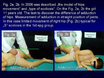
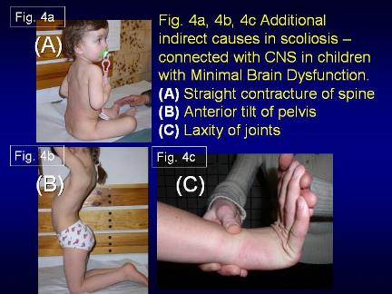
In all these cases was found the limited movement of the right hip. In some children we found an additional causes connected with the bigger or smaller disorders in brain. There were children with Minimal Brain Dysfunctions (MBD) and they had: a. primary extension contracture of the trunk, b. anterior tilt of the pelvis and c. laxity of the joints.
Biomechanical etiology
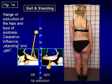
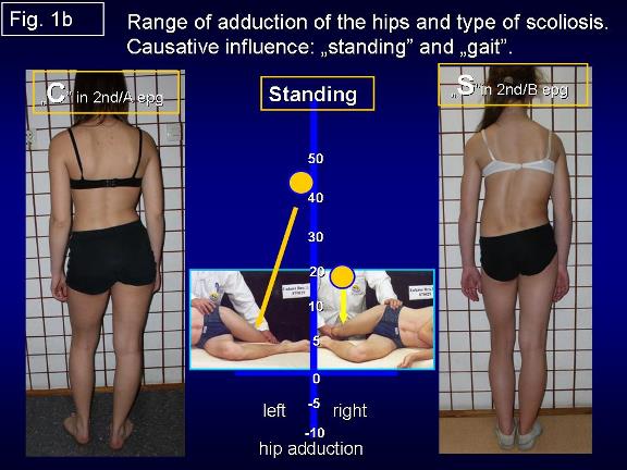
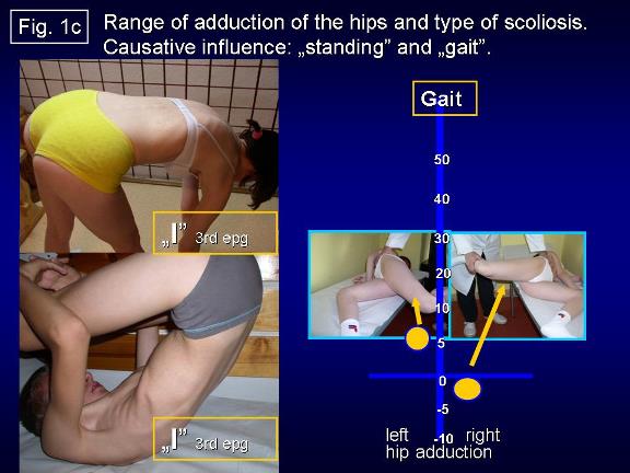
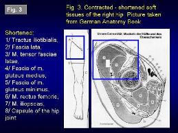
The cause of etiology of scoliosis was secret over many years [1-41]. In years 1984-2007 was found that the primary cause in etiology of scoliosis-is the asymmetry of hip movements-limited movements of right hip. In Karski T. [9] was described a specific model of hip movements-three groups of asymmetries-leading to four types of scoliosis. Next important cause in development of scoliosis is function: gait and standing on the right leg (Figure 1a, Figure 1b and Figure1c). In figures 2 and 3 is presented the test of adduction and anatomy of shortened soft tissue (Figure 2, 3).
The asymmetry of movements of the hips is one of the symptoms of Syndrome of Contracture (SofC) according to Prof. Hans Mau-Tübingen, Germany (in German Siebenersyndrom) [18]. Since 2006 we talk in Lublin about the Syndrome of Contracture and Deformities (SofCD) because we add the varus deformity of the shanks in newborns and babies as the eighth deformity.
This varus deformity, in certain conditions-may lead to Blount disease in older children [18,20-23]. In development of scoliosis the additional secondary influences can come from Central Nerve System (CNS) in children with MBD and examples are presented in (Figure 4a, Figure 4b, and Figure 4c).
Classification
Three groups and four types of scoliosis (Figure 1a, Figure 1b and Figure 1c):
(1) Scoliosis S 1st Etiopathological Group (epg)-3D. Double curve. Stiff spine. Rib hump on the right side of the thorax. Specific model of hips movements. Connection with gait and permanent standing at ease on the right leg. Beginning of deformity in 2-3 years of life. Clinical symptoms appears of the age of 5-6 years.
(2a) Scoliosis C 2nd/A epg-1D or 2D. One curve-lumbar left convex. Spine flexible. Specific model of hips movements. Connection with permanent standing „at ease on the right leg. Beginning of deformity at the age of 2-3. Clinical symptoms appear at the age of 8-10.
(2b) Scoliosis S 2nd/B epg-2D or 3D. Two curves-lumbar left convex and thoracic right convex. Specific model of hips movements. Connection with permanent standing at ease on right leg and additionally with laxity of joints or/and harmful in previous incorrect therapy/exercises. Beginning of deformity at the age of 2-3. Clinical symptoms appear of the age of 10-12 years. In the 2nd/A and 2nd/B types of scoliosis - the spine is flexible.
(3) Scoliosis I 3rd epg-2D or 3D. Specific model of hip movements. Deformity has the form of a stiff spine. No curves or small ones. The cause is gait only. Such spine deformity was until 2004 never included and classified as scoliosis. Beginning of deformity at the age of 2-3. Clinical symptoms are stiffness of the spine in children and in adults permanent pain. Why stiffness? In situations of maximal limited adduction, internal rotation and very often also extension of the right hip-appears compensatory movement in the pelvis and in the spine with every step during gait. This rotation movement in inter-vertebral joints is bigger than normal and has the character of distortion and in result causes fibrosis and stiffness.
Previous therapy
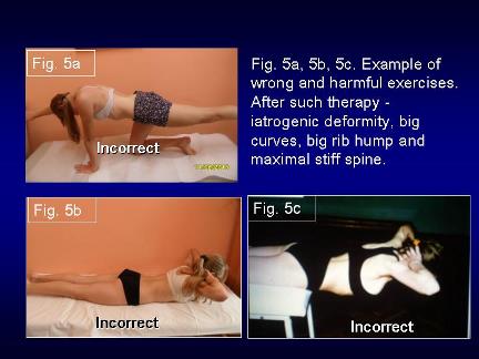
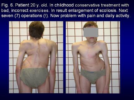
The various extensions exercises to receive strong muscles were and are only wrong and improper. They cause iatrogenic deformity of the spine-bigger curves, bigger rib hump and more stiff spine. The children after such therapy very often need surgery. The bad results after improper therapy were explained as the natural history of scoliosis (Figure 5a, Figure 5b, Figure 5c and Figure 6).
New, proper therapy
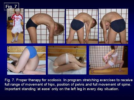
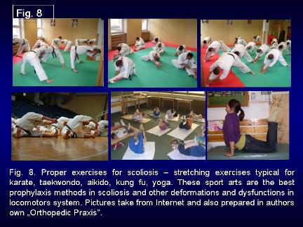
Only stretching exercises giving symmetry of movements and next symmetry of growth and the development of the pelvis and the spine are proper Figures: 7, 8. The best are stretching exercises like karate, taekwondo, aikido, kung fu and others (Figure 7 and Figure 8).
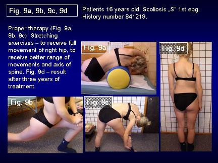
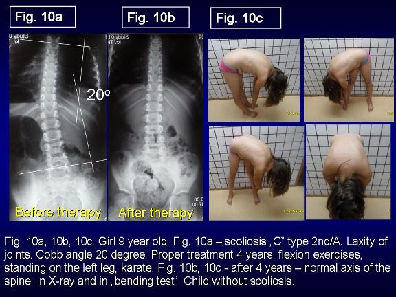
Very important are flexion-rotation exercises for spine. Such treatment gives good results (Figure 9a, Figure 9b, Figure 9c, Figure 9d, Figure 10a, Figure 10b). In this part of the paper it is my ethical obligation to inform-that-flexion exercises for the spine in scoliosis in Poland many years ago (1960-1980) were introduced by Prof. Malawski S [27]. But at this time the biomechanical influence going from the hips, pelvis and the factors: standing at ease on the right leg and gait was not discovered.
The possibility of causal prophylaxis
To find the danger of scoliosis we should use not only old but also the new tests:
· We should define the manner of the standing at ease-left: right leg.
· Use the test of adduction of both hips.
· Adams-Meyer test-other words-bending test for scoliosis.
· Lublin test-side bending test for scoliosis.
· Elly-Duncan test to discover the anterior tilt of pelvis and hiperlordosis of lumbar spine-due to flexion contracture of hips [8].
· Kneeing test-to discover the anterior tilt of pelvis and hiperlordosis of lumbar spine.
Rules of prophylactic recommendations against scoliosis:
· Standing at ease only on the left leg.
· Sitting relax-never straight up.
· Sleeping in embryo position.
· Active participation in sport in school and additionally in clubs-the best are karate, kung fu, taekwondo, aikido, yoga.
· Physiotherapy/Kinesio-therapy to obtain full, symmetrical movement of both hips and movement of the spine-flexion, deviation, rotation. Especially important is to recover the full adduction and internal rotation movement of the right hip.
Discussion and my remarks
Information to these problems is presented on the Website www.ortopedia.karski.lublin.pl from 2006 [41]. In Poland, in 1995-2009 I have given many lectures about the problem of scoliosis-at Polish Orthopedics and Traumatology Congresses in Lodź, Szczecin, Poznan, but till now the explanation of etiology and the rules of new therapy is not understood nor accepted. Why-because the conviction that the scoliosis is idiopathic is so deeply incrusted in the minds of many doctors, professors-that nobody searches for new knowledge.
My lectures have also been presented abroad in The International Research Society of Spinal Deformities (IRSSD) Meetings in Athens (2002), Genth (Belgium 2006), Liverpool (UK-2008) and in Poznan (Poland 2012) as well in Kolobrzeg (Poland 2009) during Scoliosis Course Meeting of SICOT and in Czech Republic during SICOT Meeting in 2011 and twice at the Society on Scoliosis Orthopedic and Rehabilitation Treatment (SOSORT) Congresses in Athens and in Wiesbaden and remain till now without any response. Only Professor Martha Hawes [32] and Professor Jan Stokes [35,36] from the USA, as well Professor John Sevastik [37,38] and Dr. Helen Normelly [33] from Sweden, Professor Stefan Malawski [27,28] and Professor Kazimierz Rąpała [34,40] from Poland, as well Professor Harald Thom [39] from Germany-understood my explanation of the biomechanical etiology of the so-called idiopathic scoliosis.
In the article there is all the information about etiology, classification, new therapy, but-the most important-the rules of causal prophylaxis of scoliosis. The conviction that can be other etiology of idiopathic scoliosis-for example presented in articles of Milan Roth (Czech Republic) or divagations of Prof. Mikhail Dudin from St. Petersburg- Russia (my friend, we had many personal discussions) not give the answer to all questions about properties of scoliosis. Only biomechanical etiology answers all questions and because of this we speak so-called idiopathic scoliosis. The biomechanical etiology is confirmed over many years by some group of scientist but not known in all counties in the world. Children of the world are waiting for prophylaxis. I hope that from Illinois, USA, the knowledge will spread to other countries, including Poland.
Conclusions
The biomechanical etiologies of the so-called idiopathic scoliosis explain all questions connected with this spine deformity. Development of scoliosis and the types of spine deformity are connected with pathological model of hips movements-limited movements of right hip and the function- standing at ease on the right leg and gait [9]. Restricted range of movements in the right hip is one of the symptoms of the Syndrome of Contractures and Deformities according Prof. Hans Mau and Lublin observations [18].
Every type of the scoliosis starts to develop at the age of 2-3. If start to be scoliosis-the development is slowly, over years, long time secret for parents and doctors if they do not use the new tests. The infantile scoliosis in not the So-Called Idiopathic Scoliosis. The causal prophylaxis of scoliosis is possible and should be introduced in every country. Start for prophylaxis in age of 4-6 years.
The rules of prophylaxis for all children are:
· Standing at ease on the left leg.
· Sitting in a relaxed position.
· Sleeping in an embryo position.
· Active participation in sports in school and at home every day.
· Especially beneficial sports are: karate, taekwondo, aikido, kung fu and other similar.
The special message to all doctors, to all nurses-tray to understand the new knowledge and proof/check their truest. Never say-no in the first moment. Even anecdotal cause-standing at ease on the right leg-words of my friends-from Europes Country-is in the first place-real and true. Please remember-standing at ease on the right leg is true cause in etiology of the so-called idiopathic scoliosis.
Acknowledgement
I would like to express my many thanks to David Poynton and Honorata Menet for correction of the article.
References
1. Burwell G, Dangerfield PH, Lowe T and Margulies. Etiology of adolescent idiopathic scoliosis: current trends and relevance to new treatment approaches (2000) J Spine 14: 324
2. Dangerfield PH, Dorgan JC, Scutt D, Gikas G and Taylor JF. Stature in Adolescent Idiopathic Scoliosis (AIS) (1995) 14 Meeting EPOS, Belgium, pp- 210.
3. Green NE and Griffin PP. Hip dysplasia associated with abduction contracture of the contralateral hip (1982) J Bone & Joint Surgery 64: 1273-1281. https://doi.org/10.2106/00004623-198264090-00002
4. Tylman D. Patomechanika bocznych skrzywień kręgosłupa, Wydawnictwo Severus (1995) Wydawnictwo Severus, Poland.
5. Heikkilä E. Congenital dislocation of the hip in Finland. An epidemiologic analysis of 1035 cases (1984) Acta Orthop Scandinavica 55: 125-129. https://doi.org/10.3109/17453678408992322
6. Hensinger RN. Congenital dislocation of the hip (1979) Clinical Symp 31: 270
7. Howorth B. The etiology of the congenital dislocation of the hip (1977) Clin Orthop 29: 164-179.
8. Karski T. Etiology of the so-called idiopathic scoliosis. Biomechanical explanation of spine deformity. Two 272 groups of development of scoliosis. New rehabilitation treatment. Possibility of prophylactics, Studies in 273 Technology and Informatics (2002) Res into Spinal Deform 91: 37-46.
9. Karski T, Kalakucki J and Karski J. Syndrome of contractures (according to Mau) with the abduction contracture of the right hip as causative factor for development of the so-called idiopathic scoliosis (2006) Stud Health Techno Inform 123: 34-39
10. Karski T. Explanation of biomechanical etiology of the so-called idiopathic scoliosis (1995-2007). New clinical and radiological classification (2010) Locomotor System 17: 26-42.
11. Karski T. Biomechanical etiology of the so-called idiopathic scoliosis (1995-2007)-connection with 279 syndrome of contractures-fundamental information for pediatricians in program of early prophylactics/280 (2011) J China Medical Science 8: 278-281.
12. Karski T. Biomechanic factors in the etiology of idiopathic dinominated scoliosis. New 282 classification. New clinical tests and new conservative treatment and prophylaxis (2010) Physiotherapy 39: 85-152
13. Karski T. Biomechanical Etiology of the So-called Idiopathic Scoliosis (1995-2007). New Classification: 285 Three Groups, Four Sub-types. Connection with Syndrome of Contractures (2010) Pan Arab J Orth Trauma 14: 286-287.
14. Karski T. Biomechanical Etiology of the So-called Idiopathic Scoliosis (1995 - 2007). Three Groups Four Types in the New Classification (2013) J Nov Physiother S2: 289-290. https://doi.org/10.4172/2165-7025.s2-006
15. Jacek K and Karski T. So-Called Idiopathic Scoliosis. Diagnosis. Tests examples of children incorrect 291 treated. New therapy by stretching exercises and results (2013) J Nov Physiother 3: 292-293.
16. Karski T. Biomechanical Aetiology of the So-Called Idiopathic Scoliosis. New Classification (1995-2007) in Connection with Model of Hips Movements (2014) Global Journal of Medical Research H: Orthopedic and Musculoskeletal System 14.
17. Karski T. Biomechanical etiology of the so-called idiopathic scoliosis (1995-2007) connection with „syndrome of contractures-fundamental information for pediatricians in program of early prophylactics (2014) Surgical Science 5: 33-38.
18. Karski T and Jacek k. Syndrome of Contractures and Deformities according to Prof. Hans Mau as Primary Cause of Hip, Neck, Shank and Spine Deformities in Babies, Youth and Adults (2015) Ameri Rese J Med Surgery 1: 26-35.
19. Karski T and Jacek K. Biomechanical etiology of the so-called Idiopathic Scoliosis (1995-2007). Causative role of „gait and „permanent standing at ease pn the right leg. New classification. Principles of new therapy and causal prophylaxis (2015) Canad Open Med Sci Med J 1: 1-16.
21. Karski T. Physiotherapy-Correct, or Incorrect, Based on Wrong Principles of Treatment. Example for Spine, Hip, Knee, Shank and Feet (2017) Ortho Rese Online J 1: 3-6. https://doi.org/10.31031/OPROJ.2017.01.000502
22. Karski T, Karski J, Karska K, Karska K and Menet H. pediatric prophylaxis program of motor system deformations and illnesses in children. problems of spine, hips, knees and feet (2018) EC Paediatrics 7.7: 704-714.
23. Karski T, Karski J, Karska K, Karska K and Menet H. Prophylactic rules for newborns, babies, children and adults in problems of hip, knee, shank, feet and spine (2018) Ortho Res Online J 2:110-112. https://doi.org/10.31031/OPROJ.2018.02.000530
24. Karski T and Karski J. Low back pain-problem neurological and orthopedic symptoms. Causes of treatment back pain-neurology-orthopedic problems clinic, causes, therapy and prophylaxis advances in practical Neurology (2016) Czelej Publishing House 4: 9-16.
25. Karski J and Karski T. Imperfect hips as a problem at an older age. early and late prophylactic management before arthrosis (2016) Jacobs J Physiotherapy Exercises 1: 15.
26. Karski T. Biomechanical aetiology of the so-called adolescent idiopathic scoliosis (AIS). lublin classification (1995-2007). Causative influences connected with gait and standing at ease on the right leg (2018) J Orthopedics Bone Res 1: 1-10.
27. Malawski S. Własne zasady leczenia skolioz niskostopniowych w świetle współczesnych poglądów na etiologię i patogenezę powstawania skolioz (1994) Chir Narz Ruchu i ortop Pol 59:189-197.
28. Stefan M. confirmation of Biomechanical etiology of the so-called idiopathic scoliosis (1995).
29. Mau H. Etiopathogenesis of scoliosis, hip dysplasia and torticollis of infancy (1979) J Ortho 5: 601-605.
30. Mau H. Atiopatogenesis of scoliosis, orthopedic library (1982) Enke Verlag Stuttgar 33: 296-297.
31. Hans M. confirmation of Biomechanical etiology of the so-called idiopathic scoliosis (1998)
32. Martha H. confirmation of Biomechanical etiology of the so-called idiopathic scoliosis (2002)
33. Normelly H. Asymmetric rib growth as an aetiological factor in idiopathic scoliosis in adolescent girls (1985) United Kingdom, 1-103.
34. Rąpała K. in Tylman D. Patomechanika bocznych skrzywień kręgosłupa, Wydawnictwo Severus, Warszawa, 1995, Seiten 167.
35. Stokes IAF. Studies in technology and informatics (1999) Res into Spinal Deform 59: 1-385.
36. Stokes J. confirmation of Biomechanical etiology of the so-called idiopathic scoliosis (2006)
37. Sevastik J and Diab K. Studies in technology and informatics (1997) Res into Spinal Deform 37: 300-301.
38. John S. confirmation of Biomechanical etiology of the so-called idiopathic scoliosis (2008)
39. Harald T. confirmation of Biomechanical etiology of the so-called idiopathic scoliosis (1995)
40. Tylman D. Automechanika lateral curvatures of the spines (1995) Wydawnictwo Severus, Poland.
41. www.ortopedia.karski.lublin.pl
*Corresponding author
Tomasz Karski, Department of Pediatric Orthopedic and Rehabilitation, Vincent Pol University, Poland, Tel: +48 604 933 234, E-mail: tmkarski@gmail.com
Citation
Karski T. Biomechanical etiology of the so-called idiopathic scoliosis (Adolescent Idiopathic Scoliosis [AIS]). New classification rules of therapy and prophylaxis (2019) Nursing and Health Care 4: 81-85.
Keywords
Scoliosis, Biomechanical etiology, Symptoms, New classification, Therapy, Prophylaxis.
